Comprehensive Eye Exam
Maintain the health of your eyes with annual comprehensive vision and eye health examinations. The comprehensive eye exam at You & Eyes includes the following:
- Complete internal/external health check of the eye
- Eye focusing, eye teaming, and eye movement abilities assessment
- Ocular muscles, blood vessels, and nervous systems evaluation
- Risk assessment of eye conditions resulted from general health problems and family history of eye diseases
- Eye glass prescription evaluation
- Dilation for internal health evaluation
Having a regular eye exam is crucial in protecting your and your family’s eyesight. These exams allow your doctor to detect changes in the front of your eye so alterations can be made to your eyeglass or contact lens prescription. However, your doctor also needs to look at the back of your eye, the retina, to check that it is healthy and not damaged or showing signs of disease.
Many eye diseases, if detected early, can be treated successfully.
Regular eye care can uncover both eye and systemic (entire body) problems. If these problems are left untreated, there is a risk of disability, suffering, and loss of productivity. The goals of an eye exam is to avoid or minimize adverse effects on the eye and vision, as well as to identify potential problems early in order to prevent any problems, which may potentially lead to vision loss.
There are various retinal diseases and conditions of the eye which result in loss of vision.
There are also diseases such as diabetes and high blood pressure which can be discovered by examining the eyes. Side effects of drugs may also sometimes be observed during eye examinations. Again, early detection is the key factor in treatment and sight preservation.
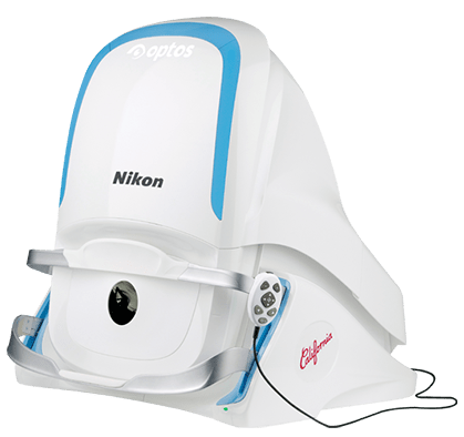
We also offer retinal imaging with the optomap ultra-widefield retinal imaging and optical coherence tomography (OCT) iWellness scanning.
(retinal imaging can be added on to your comprehensive eye exam for an additional cost)
Your retina (located in the back of your eye) is the only place in the body where blood vessels can be seen directly. This means that in addition to eye conditions, signs of other diseases (for example, stroke, heart disease, hypertension, and diabetes) can also be seen in the retina. Early signs of these conditions can show on your retina long before you notice any changes to your vision or feel pain. While eye exams generally include a look at the front of the eye to evaluate health and prescription changes, a thorough screening of the retina is critical to verify that your eye is healthy.
What is an optomap Image?
Getting an optomap image is fast, painless, and comfortable. Nothing touches your eye at any time. It is suitable for the whole family. To have the exam, you simply look into the device one eye at a time (like looking through a keyhole) and you will see a flash of light to let you know the image of your retina has been taken.
Under normal circumstances, dilation drops might not be necessary, but your eye care practitioner will decide if your pupils need to be dilated depending on the health of your eyes. The image capture takes less than a half-second and they are available immediately for you to see your own retina. You see exactly what your eye care practitioner sees – even in a 3D animation.
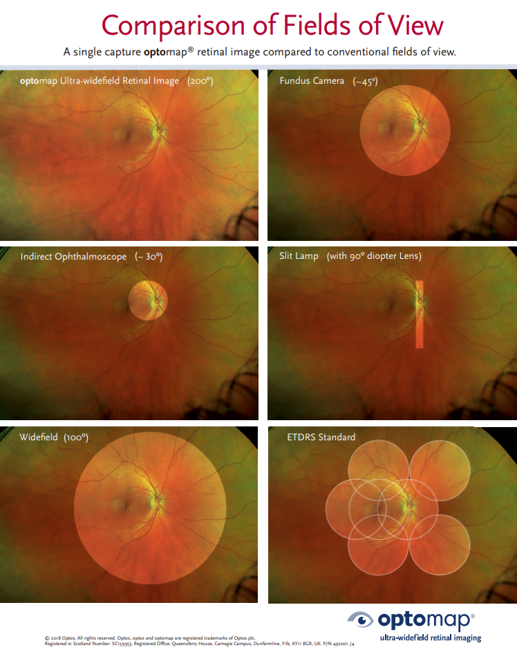
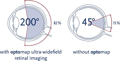
Early signs of disease can be present in the periphery of your retina and remain undetected for a long time when using traditional methods. The optomap ultra-widefield retinal image is a unique technology that captures more than 80% of your retina in a single image while traditional imaging methods typically only show 15% of your retina at one time.
Benefits of an optomap Image
The benefits of having an optomap ultra-widefield retinal image taken are:
– optomap facilitates early protection from vision impairment or blindness
– Early detection of life-threatening diseases like cancer, stroke, and cardiovascular disease
The unique optomap ultra-widefield view helps your eye care practitioner detect early signs of retinal disease more effectively and efficiently than with traditional eye exams.
Early detection means successful treatments can be administered and reduces the risk to your sight and health.
Why is a retinal exam so important?
Some of the first signs of diseases such as stroke, diabetes and even some cancers can be seen in your retina, often before you have other symptoms. An optomap makes it easier to see them. It may help your eye doctor detect problems more quickly and easily. Unlike traditional retinal exams, the optomap image can be saved for future comparisons. Optomap images are created by non-invasive, low-intensity scanning lasers. No adverse health effects have been reported in over 150 million sessions.
Is an optomap safe for children?
Yes. In fact, many vision problems begin in early childhood, so it’s important for children to receive quality routine eye care.
What is Optical Coherence Tomography (OCT)?
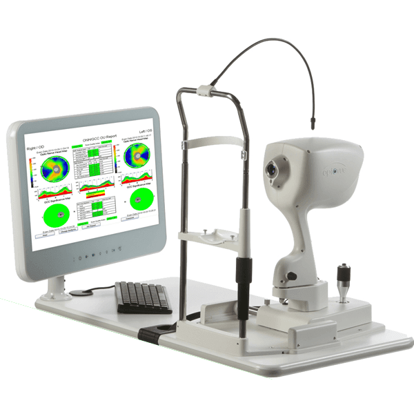
Optical coherence tomography (OCT) is a non-invasive imaging test that uses light waves to take cross-section pictures of your retina. With OCT, your Optometris can see each of the retina’s distinctive layers. This allows your doctor to map and measure their thickness. These measurements help with diagnosis and they also guide treatment for glaucoma as well as retinal disease, like age-related macular degeneration (AMD) and diabetic eye disease.
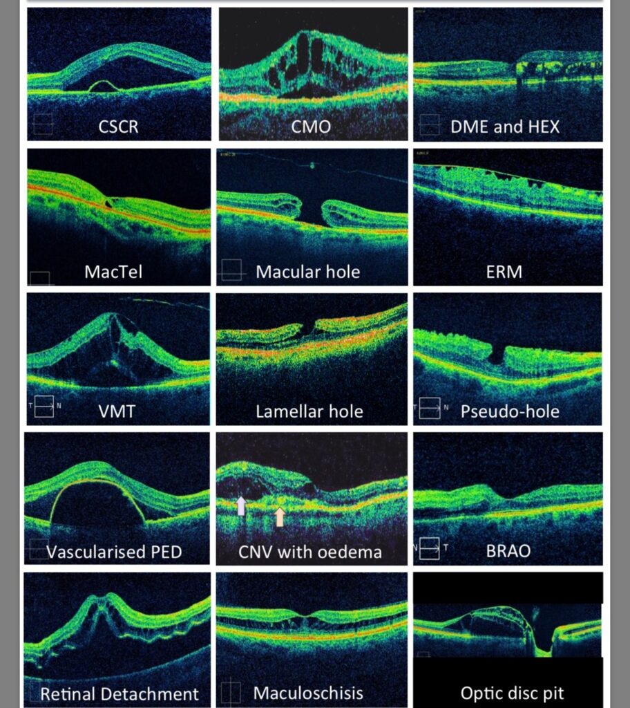
What Conditions Can OCT Help Diagnose?
OCT can help diagnose many eye conditions, including:
- macular hole
- macular pucker
- macular edema
- age-related macular degeneration
- glaucoma
- central serous retinopathy
- diabetic retinopathy
- vitreous traction
- abnormal blood vessels
- blood vessel blockage
OCT is often used to evaluate disorders of the optic nerve as well. The OCT exam helps your optometrist see changes to the fibers of the optic nerve. For example, it can detect changes caused by glaucoma.
OCT relies on light waves. It cannot be used with conditions that interfere with light passing through the eye. These conditions include dense cataracts or significant bleeding in the vitreous.
Schedule an appointment with You & Eyes, and we will be in touch with you shortly!